Introduction
Surgical procedures have evolved from purely mechanical dissection relying on cold‑steel scalpels to sophisticated techniques that utilize complex energy modalities for tissue cutting and hemostasis, marking a major leap in the field of surgery. In modern operating rooms, energy‑based surgical devices have become indispensable core tools. Interacting with biological tissues via diverse physical principles, they enable precise, efficient and safe surgical operations. Rooted in biophysical fundamentals, this paper analyzes the core working mechanisms of five mainstream surgical energy platforms: monopolar electrosurgery, bipolar electrosurgery, argon beam coagulator, ultrasonic scalpel, and cavitational ultrasonic surgical aspirator (CUSA). It explains how each technology converts electrical or mechanical energy into targeted tissue effects, and correlates their physical properties with clinical application scenarios, advantages and limitations.
Section 1: Fundamental Physics of Electrosurgery in Biological Tissues
1.1 Necessity of High‑Frequency Alternating Current
One of the core functions of electrosurgical units (ESUs) is to convert standard mains electricity (50‑60 Hz) into current at far higher frequencies, typically ranging from 100 kHz to 5 MHz¹. This frequency conversion constitutes the cornerstone of electrosurgical safety. At low frequencies (e.g., 60 Hz), rapid potential fluctuations of electric current trigger repeated depolarization of nerve and muscle cell membranes, resulting in uncontrolled muscle spasm, severe pain, and even life‑threatening arrhythmias⁴. By contrast, when the current frequency exceeds 100 kHz, the polarity alternates so rapidly that voltage‑gated ion channels on cell membranes cannot respond to elicit action potentials². Under such circumstances, intracellular ions merely oscillate rapidly in‑situ under the electric field and generate heat through friction, without evoking neuromuscular excitation.
This principle allows engineers to exploit the thermal properties of electric current rather than its electrical‑stimulus characteristics. Such transformation redefines electrosurgery as a technique that employs electric energy as a precise, controllable heat source, instead of "electrically shocking" tissues. Energy is delivered in a form “invisible” to the neuromuscular system, enabling thermal treatment of tissues without physiological electrical interference.
1.2 Joule‑Heating Effect: The Driver of Tissue Responses
The fundamental principle of electrosurgery lies in the conversion of electrical energy into thermal energy⁷. As electric current passes through biological tissue, the tissue offers resistance (or impedance under alternating‑current conditions) to current flow, which generates heat⁸. The amount of heat (Q) produced follows Joule's Law, whose mathematical expression is:
Q=I2Rt
where denotes current intensity, denotes tissue resistance, and denotes the duration of current application⁷.
Joule’s Law serves as the core equation for electrosurgery. It reveals that the heat generated within tissue does not have a linear relationship with current intensity, but instead follows a quadratic relationship — doubling the current intensity quadruples heat production. This accounts for the rapid and intense thermal effects achievable with electrosurgery. Tissue impedance (R) represents another critical variable. Water‑ and electrolyte‑rich tissues such as muscle are good conductors (low R value), whereas tissues including fat and bone are poor conductors (high R value)¹². As tissues undergo desiccation and coagulation during heating, their impedance rises sharply, which in turn restricts further current flow⁴.
This phenomenon gives rise to a vital intrinsic negative‑feedback mechanism: the self‑limiting nature of the coagulation process. According to Ohm’s Law (I=V/R), with constant voltage (V), a substantial increase in tissue impedance (R) causes a sharp drop in current (I). Since heat production is proportional to the square of current, the heating effect diminishes markedly once tissue desiccation occurs. This explains why electrocoagulation is generally depth‑self‑limited: the desiccated outer tissue layer effectively acts as an insulator, protecting deeper tissues from electric‑current exposure.
1.3 Current Density: The Dominant Variable Determining Tissue Effects
Specific tissue effects (cutting or coagulation) depend on the heating rate, which is primarily governed by current density (current intensity per unit area)⁴. Accordingly, Joule’s Law can be formulated more precisely as:
Heat ∝ (Current / Cross‑sectional Area)² × Resistance × Time
This concise formula represents the most important concept for understanding electrosurgical practice¹⁰. A tiny electrode tip concentrates current within an extremely small area, generating an extremely high current density. This causes an instantaneous, sharp rise in tissue temperature above 100 °C, resulting in immediate boiling and vaporization of intracellular water and explosive rupture of cells, thereby achieving tissue vaporization for cutting². In contrast, employing a larger electrode or the lateral surface of an electrosurgical blade distributes the same current across a broader region. Such lower current density produces a slower heating process (temperatures between 60‑99 °C), denaturing and desiccating tissue proteins without reaching the threshold for vaporization — this corresponds to coagulation². The return pad in monopolar surgery exemplifies minimal current density; its large surface area ensures no thermal effects are generated as current flows back to the generator⁴.
Accordingly, surgeons control tissue effects not merely by adjusting generator power (wattage), but predominantly by manipulating geometric and temporal parameters. Surgeons precisely regulate current density by selecting electrode geometry (e.g., fine needle‑shaped electrodes for cutting, or ball‑shaped electrodes for coagulation)⁷, modifying the contact area between electrode and tissue (using the tip versus the side of the blade), and controlling application duration (parameter t in Joule’s Law)¹². This demonstrates that the physical electrode‑tissue interface is equally critical as the electronic settings of the generator. When eschar accumulated at the electrode tip increases resistance, a surgeon who simply calls for “more power” instead of cleaning the electrode demonstrates a misunderstanding of this fundamental principle¹.
1.4 Role of Waveforms: Modulation of Tissue Effects
Electrosurgical generators can produce different current waveforms to achieve distinct tissue effects. A continuous, unmodulated low‑voltage sine wave is utilized for the “pure cut” mode². By contrast, an intermittent, modulated high‑voltage, highly damped waveform is applied for the “coagulation” mode². The “blend” mode combines characteristics of both.
Duty cycle is an engineering term describing the energy output pattern. It refers to the percentage of time during one full operating cycle in which the energy device actually delivers energy.
It can be visualized as a rapidly flickering light bulb:
‑ 100 % duty cycle: The bulb stays illuminated continuously without turning off. For electrosurgery, this corresponds to the “pure cut” mode. Energy is delivered steadily to tissue, causing an instantaneous, sharp temperature rise and rapid boiling‑vaporization of intracellular water, which yields clean cutting performance.
‑ Low duty cycle (e.g., 6 %): The bulb lights only for a brief instant and then remains off for a long interval, repeating this cycle. In electrosurgery, this matches the “coagulation” mode. Energy is delivered in short pulses. During the “off‑time” (the remaining 94 % of the cycle), generated heat has sufficient time to dissipate into surrounding tissues rather than concentrating at a single spot. This slow, gentle heating denatures tissue proteins and constricts blood vessels to achieve hemostasis (coagulation), yet does not reach the threshold for tissue vaporization. The higher voltage required in coagulation mode serves to overcome the progressively rising impedance of desiccating tissue and enables current to jump across tiny gaps to the tissue via electric spark, known as fulguration².
‑ Blend mode: The duty cycle falls between the above‑mentioned modes, for example delivering energy for 50 % of the cycle and pausing for the other 50 %. This enables tissue cutting while providing a certain degree of hemostatic effect.
Accordingly, duty cycle is a key physical parameter employed by electrosurgical generators to precisely modulate and control tissue responses (cutting, coagulation, or a combination of both). By adjusting the duty cycle, surgeons can strike a trade‑off between cutting efficiency and hemostasis to accommodate diverse surgical requirements. Efficient cutting demands highly concentrated energy to vaporize cells with minimal lateral thermal injury. Effective hemostasis, by contrast, requires slower, more extensive heating to induce vasoconstriction and protein denaturation. Therefore, the “cut” waveform prioritizes efficiency at the cost of partial hemostatic performance; the “coagulation” waveform prioritizes hemostasis yet causes greater lateral thermal injury². The “blend” mode attempts to achieve a balance between the two by tuning duty cycle and voltage.
Section 2: Monopolar Electrosurgical System
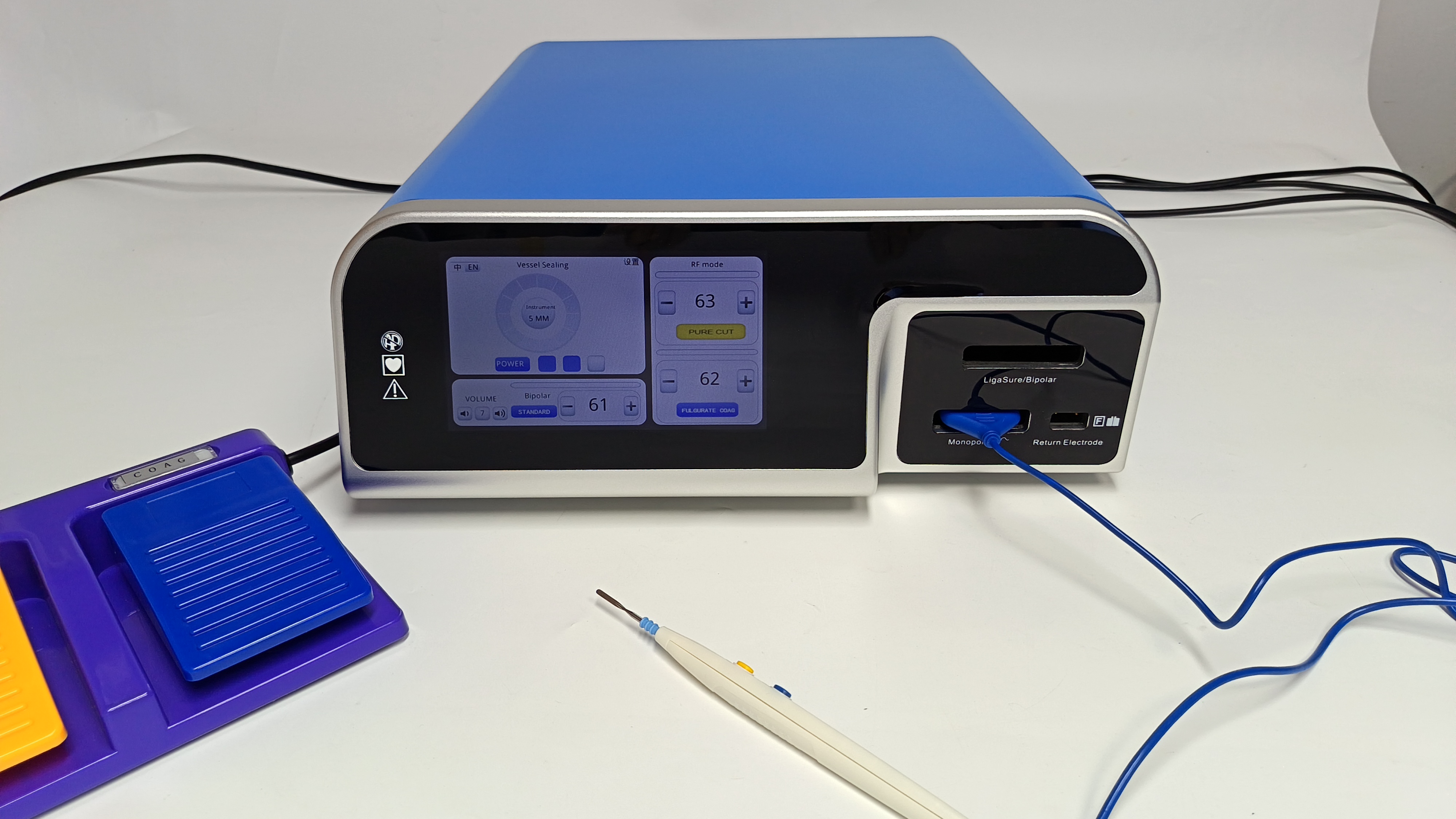
Figure showing the monopolar electrosurgical unit.
2.1 Working Principle and Complete Electrical‑Circuit Path
In monopolar mode, the electric‑current loop consists of the electrosurgical generator, the active electrode (e.g., electrosurgical pencil), the patient’s body, and a large‑area return pad (also referred to as dispersive electrode or return electrode). Current ultimately flows back to the generator through the return pad to form a closed circuit⁴. The entire patient’s body lies between the active electrode and the return pad⁴.
The term “monopolar” is in fact a conventional misnomer, since every circuit requires two electrodes to complete a loop. This nomenclature indicates that the surgical effect is concentrated on the single “active” electrode at the operative site⁴. Its core design principle lies in the vast difference in current density between the active electrode and the return pad: extremely high current density occurs at the active electrode and produces intense thermal effects, whereas the large surface area of the return pad yields very low current density and consequently no thermal injury⁴. This design enables surgeons to deliver energy via a small, precise instrument to any body region, provided that the current‑return path remains safe and intact.
Treating the patient as an integral component of the circuit also introduces inherent risks. Given that current must traverse a substantial portion of the patient’s body, the system is vulnerable to “current diversion” or alternative‑site burns. If current discovers a return path with lower resistance than the intended return pad — for instance, via electrocardiogram (ECG) electrodes or contact points with metallic operating‑table components — current will take this alternative route¹⁰. Should the contact area of such an alternative path be small, current density at that location may rise sufficiently high to cause severe burns at sites remote from the surgical field. This inherent hazard is a direct consequence of the physical characteristics of the monopolar electrosurgical circuit.
2.2 In‑depth Discussion of Modes and Tissue‑Induced Effects
Monopolar devices can produce four primary tissue effects:
1. Electrosection: Continuous low‑voltage current vaporizes tissue at the electrode tip to create an incision².
2.Desiccation / Contact Coagulation: The electrode makes direct contact with tissue; intermittent waveforms produce slow heating and tissue dehydration¹⁸.
3.Fulguration / Non‑Contact Coagulation: A small gap is maintained between electrode and tissue. The high voltage of coagulation mode ionizes the air in‑between to generate electric sparks. Current is transmitted to tissue through these sparks, producing superficial carbonization and coagulation over a relatively broad area².
4.Coaptation: Vessels are grasped with metal forceps. The active electrode is then applied to the forceps, and current travels through the forceps into the vessel to achieve vessel closure²¹.
These distinct effects are all achieved by manipulating the same core physical principles: waveform, voltage, and current density. From a physical perspective, fulguration merits particular attention. Its high voltage ionizes air between electrode and tissue, building a conductive path that allows current to “jump” across the gap. Though less precise than contact desiccation, this modality is highly effective for managing diffuse, widespread oozing².
The versatility of monopolar electrosurgery stems from its ability to harness multiple physical phenomena. It achieves desiccation via resistive heating, cutting through explosive vaporization, and fulguration by means of plasma discharge (electric sparks). Owing to such physical diversity, it remains the most widely used energy device in surgery to this day¹⁹. Merely by adjusting generator settings, changing electrode geometry, and varying surgical technique, a single handpiece can perform tasks ranging from skin incision to hemostasis of large wound surfaces.
2.3 Clinical Applications and Modern Safety Mechanisms
Monopolar electrosurgical units are widely used in nearly all surgical specialties, including general surgery, dermatology (for removal of warts, skin tags and skin cancers), oral surgery, and various endoscopic procedures¹⁸. To mitigate risks inherent to its circuit design, modern electrosurgical generators adopt an isolated‑from‑earth‑ground configuration and are equipped with Return Electrode Monitoring (REM) or Contact Quality Monitoring (CQM) systems¹⁰. These systems employ a split‑type return pad and continuously monitor the impedance between its two halves. If one corner of the return pad begins to peel off and the contact area changes, impedance will rise. The system immediately triggers an alarm and cuts off energy output from the generator, thus averting hazards before burns occur¹⁰.
The development of REM/CQM systems represents a direct engineering solution to the physical risk of return‑pad burns. By actively monitoring the integrity of the circuit segment with the lowest current density, these systems prevent the most common and dangerous failure modes of monopolar electrosurgery. Such technical safety safeguards bring a circuit design with inherent theoretical risks to an acceptable safety level for routine clinical practice. The history of monopolar electrosurgery reflects the co‑evolution of technology and safety. Early ground‑referenced generators carried substantial risks of alternative‑site burns, as current could leak through any grounding point¹⁰. The first innovation was the floating‑isolated generator, which confined the current loop within the generator‑patient‑return‑pad circuit. The second, more critical innovation was the REM system, which addressed the remaining major failure point: poor return‑pad contact. This clearly illustrates a trend: as technical functions and applications expand, safety systems intended to control the inherent physical risks of the design become increasingly sophisticated.
Section 3: Bipolar Electrosurgical System
3.1 Working Principle and Restricted Current Pathway
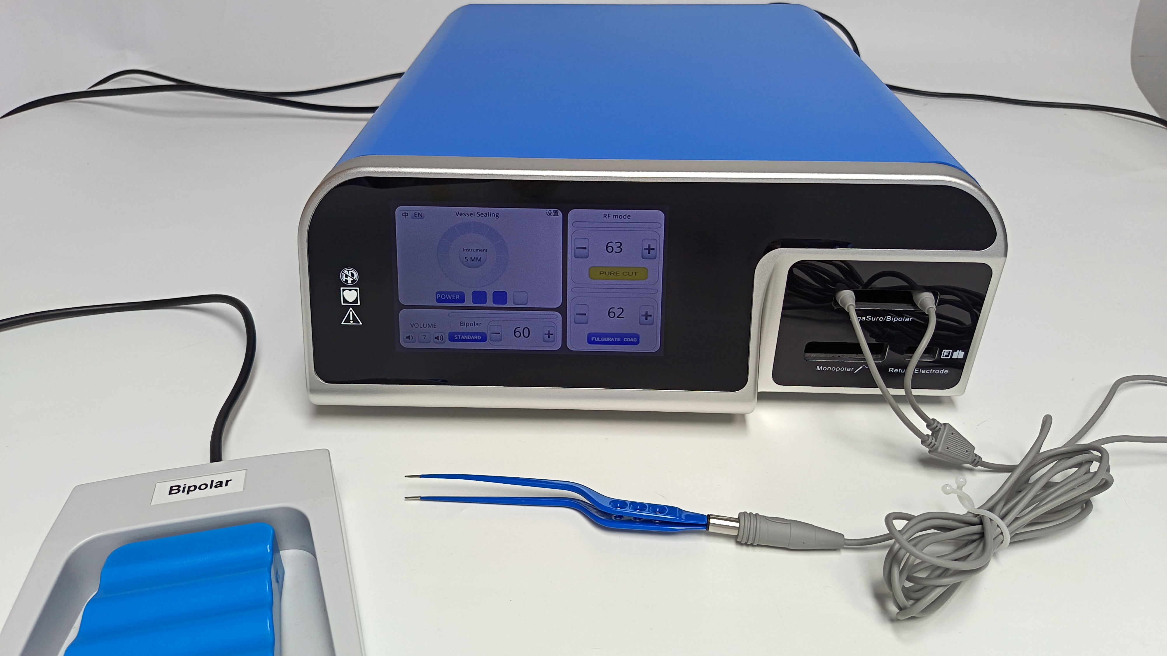
In bipolar electrosurgery, the active electrode and return electrode are integrated into a single instrument, typically the two tips of a forceps¹. Electric current is strictly confined to flow through the tissue grasped between the two electrode tips¹. Accordingly, bipolar mode does not require a separate patient return pad¹.
The physical principle behind bipolar electrosurgery is straightforward. By placing the two poles of the circuit only a few millimeters apart, the path of least resistance for current runs directly through the tiny volume of tissue held by the instrument. The rest of the patient’s body forms a far higher‑resistance pathway and is therefore excluded from the circuit. This design fundamentally eliminates the complications inherent to monopolar mode, including current diversion, alternative‑site burns, and return‑pad‑related injuries¹⁴.
The bipolar configuration fundamentally alters the risk profile by redefining the electrical circuit. Major hazards in monopolar surgery arise from the patient being part of the circuit. By contrast, the key safety feature of bipolar surgery is that the patient lies outside the circuit, with the current pathway externalized within the instrument itself. This means bipolar devices are inherently safer against stray current, making them suitable for patients with cardiac pacemakers or other electronic implants that could be disrupted by monopolar current¹⁴.
3.2 Biophysical Advantages: Precision and Reduced Thermal Spread
Bipolar surgery generally operates at lower voltages compared with monopolar coagulation mode². Since electric current is confined within a well‑defined tiny region, lateral thermal spread to adjacent tissues is minimal². This renders it an ideal option for delicate manipulations near vital structures²⁵.
Reduced lateral thermal spread is a direct consequence of the restricted current pathway. In monopolar coagulation, heat can conduct through tissues and spread far beyond the site of application. In bipolar mode, by contrast, thermal effects are almost entirely confined to the tissue sandwiched between the forceps tips. Such precision enables surgeons to coagulate small blood vessels over the surface of nerves without injuring the nerves themselves. Advanced bipolar devices (e.g., LigaSure) build upon this principle by incorporating energy‑delivery systems with pressure and feedback control. By denaturing collagen and elastin within vascular walls, they can reliably seal vessels up to 7 mm in diameter¹⁷.
The precision of bipolar energy represents more than an incremental improvement; it is an enabling technology. It substantially improves the safety of delicate procedures such as neurosurgery, ophthalmic surgery, and minimally‑invasive gynecological surgery¹. In these disciplines, achieving hemostasis without significant collateral damage is critical. The development of advanced bipolar vessel‑sealing devices has further revolutionized surgery by replacing slow and cumbersome suture ligation with a rapid, reliable sealing method, thereby shortening operative time and reducing blood loss²⁶.
3.3 Clinical Applications
Bipolar electrosurgical devices represent standard equipment for specialties demanding high precision and safety, including neurosurgery, gynecology (especially in laparoscopic and hysteroscopic procedures), urology, and ophthalmology¹. They are applied for precise coagulation, tissue sealing, and, in certain advanced modes, tissue cutting. Furthermore, they offer advantages in wet‑field surgery, as electric current will not be dispersed by surrounding fluids¹. In neurosurgery, they enable coagulation of blood vessels on the brain or spinal cord with minimal risk to neural tissue²⁵. In laparoscopic surgery, their safety features lower the risk of thermal injury to adjacent organs such as the bowel¹⁵. The capability to operate in fluid‑filled environments is essential for hysteroscopic procedures performed within the distended, fluid‑filled uterine cavity³⁰.
“Bipolar” does not constitute a single category; it covers simple low‑power forceps for delicate coagulation as well as highly engineered “advanced bipolar” devices. Rather than mere coagulators, these sophisticated systems function as “tissue‑fusion” instruments. They integrate precisely controlled pressure together with feedback loops that monitor tissue impedance to deliver optimally‑calibrated energy, denaturing and cross‑linking collagen within vascular walls to form a permanent, robust seal²⁸. This signifies a higher level of biophysical control compared with simple thermal coagulation.
Section 4: Argon Plasma Coagulation (APC)
4.1 Physical Principle of Argon Ionization
Argon Plasma Coagulation (APC) employs a stream of inert argon gas, which is ionized by high‑frequency, high‑voltage (approximately 6 kV) discharge at the probe tip to form conductive plasma¹⁰. Argon is selected because it is an inert gas characterized by chemical stability, non‑flammability, and ready ionizability¹⁰.
Plasma is the fourth state of matter. When a gas is heated to an extremely high temperature or exposed to a strong electric field, atoms lose their electrons, generating a mixture composed of positive ions and free electrons. This “ionic soup” possesses high electrical conductivity. In essence, an APC device generates a conductive argon‑plasma “beam” that forms a bridge between the probe and tissue, permitting current to flow without direct physical contact¹³. Meanwhile, the argon gas stream exerts a mechanical effect by blowing away blood and fluids from the tissue surface, allowing current to reach target tissue effectively¹⁰.
By leveraging plasma physics, APC overcomes the limitations of contact‑mode electrocoagulation for hemostasis on large, wet wound surfaces. A key drawback of conventional monopolar electrocoagulation is current dispersion within blood or saline, resulting in poor performance when managing bleeding from extensive wet wounds¹⁰. APC addresses this challenge via two mechanisms: first, the gas stream physically displaces fluids¹⁰; second, it establishes a focused conductive pathway (plasma beam) that directs current toward tissue rather than dissipating it within fluids. This represents an ingenious application of gas dynamics and plasma physics to resolve common surgical challenges.
4.2 Non‑Contact Energy Delivery and Superficial Coagulation
APC is a monopolar technique and therefore requires a patient return pad to complete the electrical circuit¹⁰. Current is conducted through the plasma beam to the tissue surface and induces rapid, superficial coagulation¹⁰. Its coagulation depth is generally shallow and self‑limiting¹³.
Self‑limitation constitutes the most critical biophysical property of APC. As current passes through and coagulates the tissue surface, the tissue desiccates and its resistance rises sharply. Seeking consistently the path of least resistance, the argon plasma beam automatically deflects toward adjacent uncoagulated tissue regions with lower resistance¹³. This means continuous APC application tends to expand the coagulated area rather than increase coagulation depth, thereby preventing deep tissue necrosis and reducing the risk of perforation¹³. The depth of injury is governed by power settings and gas flow rate, instead of application duration¹³.
The efficacy of APC relies on the substantial resistance difference between normally moist tissue and desiccated coagulated tissue. The plasma beam can be visualized as “painting” over low‑resistance zones. Once an area is “painted” (i.e., coagulated), its resistance becomes extremely high and effectively “repels” electric current. This creates a highly efficient system for managing large wound surfaces, as energy is automatically and preferentially delivered to regions still requiring treatment. Such impedance‑based targeting represents a sophisticated physics‑driven safety feature.
4.3 Clinical Applications
APC is used for rapid, non‑contact hemostasis of large wound surfaces, for example during liver surgery¹⁰. It is widely applied via endoscopy in gastroenterology to treat conditions including Barrett’s esophagus, radiation proctitis, angiodysplasia (i.e., “watermelon stomach”), and post‑polypectomy bleeding³². It is also utilized in pulmonology for ablation of endobronchial tumors³².
The clinical applications of APC align perfectly with its physical properties. Its capacity to achieve extensive, superficial coagulation makes it ideal for controlling diffuse bleeding from solid organs where deep penetration is undesirable. Its non‑contact and forward‑firing characteristics are well‑suited for endoscopic procedures, enabling treatment of lesions within the gastrointestinal tract or airways without requiring precise probe‑to‑tissue contact¹³. Its self‑limiting depth constitutes a critical safety feature within the thin‑walled gastrointestinal tract³⁴.
APC serves as a surface‑ablation tool rather than a dissecting instrument. Unlike electrosurgical knives, APC cannot perform cutting due to its overly dispersed energy density. Its sole purpose is thermal ablation of tissue surfaces. Accordingly, it acts as a complementary tool instead of a substitute for other devices. Surgeons may resect liver tumors using a scalpel or CUSA, and then employ APC to manage diffuse oozing from the exposed liver surface. This highlights the concept of multimodal surgical approaches, in which different energy devices are used sequentially at various operative stages to leverage their distinctive physical characteristics.
Section 5: Ultrasound‑Energy Systems
5.1 Ultrasonic Scalpel (e.g., Harmonic Scalpel)
5.1.1 Working Principle: Reverse Piezoelectric Effect
The handpiece of an ultrasonic scalpel system incorporates piezoelectric crystals (ceramics), which convert high‑frequency electrical signals from the generator into mechanical vibration⁴⁰. This phenomenon is known as the reverse piezoelectric effect. After amplification, these vibrations propagate along the blade, driving the blade to oscillate longitudinally at an extremely high frequency (typically 55.5 kHz, equivalent to 55 500 cycles per second)⁴⁰.
Piezoelectric materials feature a unique crystal structure that deforms upon voltage application. Rapid alternating voltage applied at the material’s resonant frequency enables efficient generation of high‑amplitude vibration. The ultrasonic scalpel represents a sophisticated acoustic system engineered to generate, amplify, and transmit these mechanical vibrations to its precisely manufactured blade tip.
The physical principles of the ultrasonic scalpel are fundamentally different from those of electrosurgery. Rather than forming an electrical circuit that includes the patient, the device functions as an acoustic system. Energy is delivered to tissue in the form of mechanical waves instead of electric current. This distinction underpins its key safety advantages: there is no risk of electrical burns, current diversion, or implant interference, and lateral thermal spread is markedly reduced⁴⁸.
5.1.2 Mechanism of Action: Triple Effects of Cavitation, Frictional Heat and Coaptive Coagulation
The ultrasonic scalpel achieves cutting and coagulation via three synchronized physical processes:
1.Cavitation: Rapid blade vibration (amplitude of 50‑100 µm) generates drastic pressure fluctuations within tissue fluid, resulting in vaporization of intracellular water at low temperatures. Formation and collapse of these bubbles disrupt and separate tissues to produce a cutting effect⁴¹.
2.Frictional Heat: Intense friction at the cellular level occurs as the blade rubs against tissue. The resultant heat denatures proteins (especially collagen) to form a viscous coagulum that seals blood vessels⁴⁴. Its operating temperature (50‑100 °C) is lower than that of electrosurgery (150‑400 °C)²⁸.
3.Coaptive Coagulation: The stationary upper jaw of the instrument clamps tissue and applies compressive force (coaptive compression), bringing vascular walls into apposition so that denatured protein coagulum forms an effective seal⁴⁴.
The sophistication of the ultrasonic scalpel lies in its ability to harness the synergistic interplay of these three effects. Surgeons can modulate the dominant effect by adjusting power, compression force and tissue tension⁴⁸. High compression and high power favor coagulation and vessel sealing, whereas high tissue tension combined with the sharp edge of the blade promotes cutting. This allows a single instrument to perform tasks that conventionally require repeated instrument changes (e.g., cutting with scissors and achieving hemostasis with bipolar electrocoagulation).
Electrosurgery seals blood vessels essentially by boiling and desiccating tissue, producing carbonized eschar. The ultrasonic scalpel operates differently. Its primary mechanism breaks hydrogen bonds within protein molecules through mechanical vibration and moderate frictional heat⁴⁴. This unwinds (denatures) proteins, which re‑form a viscous coagulum upon cooling and act as biological glue⁴⁴. This lower‑temperature, protein‑centered mechanism accounts for its reduced eschar production, less surgical smoke, and diminished lateral thermal injury compared with electrosurgery²¹.
5.2 Cavitron Ultrasonic Surgical Aspirator (CUSA)
5.2.1 Working Principle: Low‑Frequency, High‑Amplitude Ultrasonic Fragmentation
The Cavitron Ultrasonic Surgical Aspirator (CUSA) shares a similar working principle with the ultrasonic scalpel (both utilize transducers to generate ultrasonic vibration), yet it operates at a lower frequency (typically 23‑36 kHz)⁵⁴. The lower frequency enables the vibrating tip to achieve greater amplitude (longer displacement)⁵⁴. This system integrates tissue fragmentation, irrigation and aspiration functions within a single handpiece⁵⁴.
CUSA is engineered for distinct objectives: tissue emulsification and removal, rather than cutting and sealing. Its lower frequency and higher amplitude deliver a more powerful fragmenting effect, essentially acting upon tissue like a “jackhammer” at the microscopic level. The integrated irrigation function cools the tip and provides an aqueous medium for cavitation, while the aspiration function instantly removes fragmented tissue debris to maintain a clear surgical field⁵⁴.
A comparison between the Harmonic scalpel (55.5 kHz) and CUSA (23‑36 kHz) reveals a key engineering principle: tuning physical parameters to achieve divergent biological outcomes. The higher frequency of the Harmonic scalpel is well‑suited for generating rapid, localized frictional heat required for coagulation. By contrast, the lower‑frequency, higher‑amplitude setting of CUSA is optimized to maximize the mechanical destructive force of cavitation for bulk tissue fragmentation. Both are “ultrasonic devices”, but their differing operational parameters render them suitable for entirely separate surgical tasks.
5.2.2 Mechanism of Action
The primary mechanism of CUSA is selective tissue fragmentation via cavitation⁵⁴. Its vibrating tip preferentially fragments and emulsifies tissues with high water content and weak intercellular connections (e.g., hepatic parenchyma, brain tumors, adipose tissue)⁵⁴. In contrast, tissues rich in collagen and elastin with low water content (such as blood vessels, nerves, and bile ducts) are more elastic and resistant to fragmentation, and therefore remain preserved⁵⁴.
Such tissue selectivity constitutes the defining feature and most important clinical advantage of CUSA. The cavitation effect is most pronounced on inelastic, fluid‑filled cells. Elastic structures such as blood vessels can absorb and dissipate vibrational energy without rupture. This allows surgeons to “dissolve” hepatic parenchyma while preserving the intricate network of blood vessels and bile ducts (namely “skeletonization”), which can then be ligated and divided one‑by‑one under direct vision⁶⁰. This level of precision cannot be achieved by electrosurgery or conventional scalpels.
The selectivity of CUSA does not rely on the thermal or electrical properties of tissue, but on the intrinsic mechanical characteristics of tissues — particularly water content and elasticity. It exploits the physical differences between fragile tumor tissue and tough vascular tissue. This represents a higher order of surgical precision: rather than merely creating a fine incision, it selectively removes one tissue type while preserving another within complex, interwoven anatomical structures. This physical principle renders CUSA an indispensable tool in complex neurosurgical and liver resection procedures⁵⁴.
Section 6: Comparative Analysis and Clinical Application
6.1 Comparison of the Three Energy Modalities
The energy devices discussed in this report can be categorized into three fundamentally distinct energy forms:
1. Electrosurgical energy (monopolar, bipolar): Heat is generated by high‑frequency current passing through tissue resistance (Joule effect). Its effects are thermal and determined by current density and waveform.
2. Plasma‑mediated energy (argon plasma coagulation): A specialized form of monopolar electrosurgery that employs ionized gas to conduct electric current for non‑contact superficial thermal coagulation.
3. Mechanical energy (ultrasonic scalpel, CUSA): Electrical energy is converted into high‑frequency mechanical vibration. Its effects are predominantly mechanical (cavitation, tearing), with secondary thermal effects (frictional heat).
This classification facilitates understanding of these devices based on their physical interactions with tissue, which underpin all clinical differences.
6.2 Key Performance Indicators
The following two tables correlate physical principles with clinical performance, providing a clear framework for device selection.
Table 1: Summary of Physical Principles and Mechanisms of Action
Device | Primary Physical Principle | Energy‑Conversion Pathway | Primary Tissue Interaction |
Monopolar Electrosurgery | Joule heating effect, current density | Electrical energy → Thermal energy | Resistive heating, vaporization, protein denaturation |
Bipolar Electrosurgery | Joule heating effect (restricted current pathway) | Electrical energy → Thermal energy | Resistive heating, protein denaturation (tissue fusion) |
Argon Plasma Coagulation (APC) | Plasma conduction, Joule heating effect | Electrical energy → Plasma → Thermal energy | Non‑contact resistive heating, superficial coagulation |
Ultrasonic Scalpel | Reverse piezoelectric effect, mechanical vibration | Electrical energy → Mechanical energy → Thermal energy | Cavitation, frictional heat, protein denaturation |
CUSA | Reverse piezoelectric effect, mechanical vibration | Electrical energy → Mechanical energy | High‑power cavitation, tissue emulsification |
Table 2: Comparison of Clinical Performance Characteristics
Characteristic | Monopolar Electrosurgery | Bipolar Electrosurgery | Argon Plasma Coagulation (APC) | Ultrasonic Scalpel (Harmonic) | Cavitron Ultrasonic Surgical Aspirator |
Vessel‑sealing diameter | < 2 mm ²¹ | Conventional: < 3 mm ²⁸ Advanced: ≤ 7 mm ²¹ | No primary vessel‑sealing function | ≤ 7 mm (latest‑generation models) ⁶⁷ | No vessel‑sealing function |
Lateral thermal spread | Highest (>20 mm possible)⁴⁹ | Moderate (2‑4 mm)² | Superficial but potentially extensive¹⁷ | Lowest (<1‑2 mm)⁴⁹ | Extremely low (heat originates from friction) |
Surgical smoke | Highest⁵³ | Low⁵³ | Minimal²³ | Minimal (primarily water vapor)⁴⁸ | Generates liquid‑phase aerosols |
Tissue selectivity | None | None | None | None | Extremely high (preserves vessels / nerves)⁵⁴ |
Primary Clinical Functions | Cutting, coagulation, fulguration, desiccation | Precise coagulation, tissue sealing | Large‑area superficial hemostasis | Simultaneous cutting and coagulation | Tissue fragmentation and aspiration
|
Key Advantages | Versatile functionality, low cost, rapid operation | High safety, high precision, low thermal injury | Non‑contact, large‑area, superficial, effective in wet fields | Low thermal injury, minimal smoke, no electric current passing through the patient | Precise tissue selectivity, clear surgical field
|
Key Limitations | High safety risks (burn injury, current diversion) | Limited cutting capacity, relatively slow speed | Incapable of cutting, limited depth of effect | Relatively slow coagulation speed, high cost | No hemostatic capability, cumbersome operation, high cost |
6.3 Application‑Driven Device Selection: Clinical Synthesis
There is no single “best” energy device. The optimal choice relies on a thorough understanding of the biophysical principles of each device and matching them to specific surgical requirements, including tissue type, anatomical location, and surgical objectives.
● Trade‑off between speed and safety: A trade‑off frequently exists between the speed and versatility of monopolar electrosurgery and the safety and precision of bipolar and ultrasonic devices. For routine dissection in non‑critical regions, monopolar electrosurgery is fast and effective. However, near the bowel, nerves or ureters, the benefit of minimal thermal spread offered by ultrasonic or bipolar devices justifies the additional time or cost²⁷.
● Choice between hemostasis and dissection: Certain devices excel at hemostasis (e.g., advanced bipolar instruments, argon plasma coagulation), whereas others are optimized for dissection (e.g., CUSA, monopolar cutting). The ultrasonic scalpel is unique in attempting to achieve both functions simultaneously; nevertheless, even it has setting modes biased toward one of the two roles⁵¹. Selection is often determined by the vascularity of the tissue and the primary goal of the given surgical step.
● Cost‑benefit analysis: Advanced energy devices are substantially more expensive than standard monopolar and bipolar instruments⁵⁰. A key consideration for surgeons and hospitals is whether their benefits — such as reduced blood loss, shorter operative time, and potential shortening of hospital stay — justify the increased costs²⁹. The available evidence is often mixed and procedure‑specific, which complicates decision‑making.
Conclusion
Advancements in modern surgery are inextricably linked to the development of energy‑based surgical instruments. From electrosurgery leveraging the Joule heating effect, to argon plasma coagulation based on plasma physics, and ultrasonic systems employing mechanical vibration, each technology represents a sophisticated application of specific biophysical principles. Monopolar electrosurgery serves as a fundamental tool owing to its versatility and cost‑effectiveness, yet its inherent circuit‑related risks demand strict safety protocols. By confining current pathways, bipolar electrosurgery delivers unparalleled precision and safety, establishing it as the gold standard for delicate procedures. Argon plasma coagulation provides a unique non‑contact solution for extensive superficial bleeding. The ultrasonic scalpel achieves cutting and coagulation with low thermal injury via mechanical energy, while CUSA elevates tissue selectivity to new heights, enabling meticulous dissection within complex anatomical structures.
Ultimately, the surgeon’s toolbox does not consist of a single universal instrument, but rather a portfolio of high‑technology tools, each with distinct physical properties and clinical roles. A thorough understanding of the physical principles underlying these devices forms the cornerstone for individualized, precise and maximally safe surgical outcomes, and also acts as the driving force for future surgical‑technology innovation.





 苏公网安备32011802010375号